Ultrasound medical imaging equipment has experienced more than half a century of development. Especially since the 1990s, with the rapid development of medicine, mechanical materials, computer and electronic engineering technology, the performance of ultrasonic diagnostic instruments has been continuously improved, functions have been continuously improved, and applications have been expanded. . At present, no hospital can be separated from ultrasound imaging diagnostic technology. Ultrasound imaging diagnosis has high spatial resolution, high soft tissue contrast, real-time rapid imaging, simple operation method, no contraindication, no damage, repeatability, portability and economy. Together with CT, MRI and isotope imaging, it constitutes the four major imaging diagnostic techniques essential in clinical medicine.
Overview of the application of ultrasound imaging diagnostic technology in medicineThe application of ultrasound imaging diagnostic technology in medicine began in the middle of the last century. It only began to use the A-type ultrasound system to detect the thickness of isolated organs and explore some clinical disease diagnosis. Then it used M-mode ultrasound to detect normal people and The heart of patients with rheumatic heart disease; until the early 1970s, B-mode ultrasound imaging technology, which can show changes in morphological changes of organs and lesions, was applied to the clinic, and a new one of the two-dimensional section ultrasound imaging technique of organs was opened. page. In the mid-1980s, the color Doppler ultrasound diagnostic apparatus was introduced. Since it can display the dual information of the morphological structure and hemodynamic changes of organs and lesions, it also advances the level of ultrasonic imaging diagnostic technology. Until the widespread application of computer digital technology in the 1990s, the successful research of medical ultrasound three-dimensional imaging technology has made ultrasonic imaging diagnostic technology enter a higher level and a new stage of development. That is to say, from the end of the last century to the beginning of this century, the development of ultrasonic imaging diagnostic technology is amazing, and it has achieved many major technical breakthroughs. Throughout the development of ultrasound imaging diagnostic technology, it is a development process from "point" (A-type ultrasound) → "line" (M-mode ultrasound) → "face" (two-dimensional ultrasound) → "body" (three-dimensional ultrasound) It is a development process from one-dimensional array to two-dimensional array to three-dimensional array; it is a development process from static imaging to real-time dynamic imaging; it is a development process from single-parameter diagnostic technology to multi-parameter diagnostic technology; It is also a process of developing molecular images from anatomical morphological images to anatomical functional images, metabolic images, enzymes and receptors, and gene expression imaging.
Application of Digital Technology in Ultrasound Imaging Diagnostic EquipmentThe digitization of ultrasonic diagnostic instruments, from digital scanning converters to today's all-digital ultrasound transmission, reception, and imaging processes, digital technology has been widely adopted for high-performance ultrasound imaging diagnostic equipment, such as probe new code transmission and reception technology, digital sound beam Technology, digital delay technology, dynamic apodization technology, dynamic electronic focusing, dynamic aperture technology, etc. The development and application of digital technology has also promoted and promoted the high performance, intelligence and miniaturization of ultrasound imaging diagnostic equipment. The high-performance ultrasound imaging diagnostic device can not only meet the various needs of clinical disease diagnosis, but also deepen relevant basic theories and clinical medical research, thereby further promoting the ultrasound imaging diagnostic technology from simple morphology to morphological physiology and function and molecular. The development of imaging direction. Intelligent operation can realize one-button operation, such as one-button multi-function, which can adjust TGC, receiving gain, dynamic range, adjustable speed scale, Doppler baseline and many other parameters, thus avoiding complicated and cumbersome adjustment operations during inspection. . The miniaturization of the ultrasonic instrument under the premise of ensuring the required functions, the device structure is simple, such as the size of the notebook computer, whether it is bedside inspection or on-site emergency inspection or emergency field inspection, it can reflect the important clinical status of ultrasound imaging diagnostic technology and Value, at the same time, has broadened the scope of clinical application of ultrasound diagnostic technology. In addition, with the rise of the information superhighway, the wide application of communication and network technology, the ultrasound imaging diagnostic equipment of different manufacturers and different models are equipped with DICOM3.0 standard interface. In the DICOM 3.0 standard, it not only covers data dictionary, information interaction, network communication, media storage and file format, display printing, management and other aspects directly related to medical imaging, but also gradually covers the entire medical environment. Trends in capacity and data exchange. That is to say, the ultrasound imaging diagnostic equipment can be integrated into the hospital image management and communication system (PACS) together with the ultrasound imaging workstation, and even integrated into the information system of the entire hospital.
Development of Ultrasound Imaging Diagnostics Probe TechnologyThe probe, also known as the transducer, is one of the most important components in the ultrasound imaging diagnostics. Its main function is to transmit ultrasound echo signals to the human body before receiving ultrasound echo signals from the human body. High-performance, high-quality probes are not only a prerequisite for obtaining high-quality images, but also a technical guarantee for various new ultrasound imaging methods. The structure of the probe is generally composed of a main body, a casing and a wire, wherein the piezoelectric material (wafer) is the core of the body. From single-chip and multi-wafer development to tens, hundreds, or even thousands of wafers, the number of probe elements consisting of several wafers connected in parallel is expanding. At present, the main development trend of the probe is new materials, new processes, multi-array (high density), high frequency, wide frequency band and dedicated. New materials: mainly including composite materials and organic thin film materials; new technology: the piezoelectric ceramics and high polymers are combined according to a certain connection mode, a certain volume ratio and a certain spatial geometric distribution, with high sensitivity and low resistance. Advantages of resistance (matching with human tissue) and low mechanical quality factors (favorable for band broadening); High density: 1 dimension (256 arrays), 1.5 dimensions (8 &TImes; 1 2 8 array elements) 2D (60&TImes; 60 arrays); High frequency: 3MHz - 7MHz frequency probe for diagnosis of abdominal and heart diseases, 10MHz-15MHz frequency probe for superficial organ inspection, 20MHz-40MHz frequency probe Ultrasound imaging of the eye and skin, and probes of 100MHz-200MHz frequency are mainly used for ultrasound microscopy; Broadband: Broadband refers to the upper and lower limits of the operating frequency of the transducer, which enables the transmission from shallow to deep when using a probe inspection. And receiving ultrasonic echo signals of different frequencies from high to low, and at the same time, it is also an important guarantee for realizing frequency domain composite imaging, harmonic imaging and other non-linear imaging new technologies; Shape, such as a dedicated esophagus, rectum, vagina, urethra, bladder, abdominal cavity, blood vessel lumen dedicated inspection probe.
Development of several new imaging techniques in ultrasound imaging diagnosis 1. Ultrasound three-dimensional imaging technology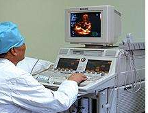
Ultrasonic three-dimensional imaging technology is a major breakthrough in the field of ultrasonic diagnostic technology. It is a new technology that is emerging in clinical ultrasound. It can obtain image information in three dimensions, thus making up for the shortcomings of two-dimensional planar imaging technology. According to the imaging principle, the three-dimensional imaging technology can be divided into static three-dimensional imaging for observing inactive organs and dynamic three-dimensional imaging and real-time three-dimensional imaging for observing cardiac morphological structures and their activities. Static three-dimensional imaging is a two-dimensional probe for rotating scanning or fan scanning scanning. A certain number of cut images are acquired into the computer for image reconstruction within a certain period of time, and the three-dimensional image of the organ is displayed. The reconstructed image is clear and the boundary is clear. The contour and depth are strong, and the morphology of organs and lesions is characteristic. It is mainly used for the presence of liquid in organs or for liquid encircling around the subject, such as liver and kidney cysts and abscesses, biliary stones and polyps, hydronephrosis and Tumors, etc.; three-dimensional image reconstruction of pancreas and duodenum can observe the stereoscopic anatomy of the head of the pancreas and surrounding tissues, and help to diagnose the lesions of the pancreatic head and common bile duct; three-dimensional reconstruction of blood vessels can realize the vascular tree without parenchymal tissue reflex The image can help to understand the vascular orientation, branching status, presence or absence of deformity, thrombosis, etc. in the organ; it also has distinctive features for the sand-like structural lesions such as ulcers, fetal facial deformities, and umbilical cord around the neck. In addition, three-dimensional ultrasound imaging can provide the doctor with the spatial location and three-dimensional shape of the tumor lesion in the body, thereby providing more accurate positioning information for ultrasound-guided interventional therapy, which helps to improve and further improve the clinical treatment effect.
With the development of high-speed scanning and sampling technology, plus the time parameters of ECG synchronization technology based on static three-dimensional imaging, dynamic three-dimensional imaging (also called four-dimensional parameters) can be realized in quasi-real-time mode; if speed is added Information, real-time 3D imaging (also known as five-dimensional parameters). Dynamic three-dimensional imaging can show the origin, location, direction and left and right relationship of large blood vessels, observe the presence or absence of defects and determine the location and shape of the defect, provide diagnosis and differential diagnosis of complex and difficult congenital heart disease; can accurately display the stereoscopic shape of the heart, Finely measure cardiac function, observe the site, extent and extent of segmental dysmotility of the wall, provide the basis for diagnosis and treatment of coronary heart disease; can display the overall structure of the valve orifice, and diagnose the stenosis and regurgitation of the valve orifice, especially the apex Valvular flap and prolapse, chordae rupture and other valve diseases have important significance; can also display stereoscopic dynamic images of intracardiac blood flow, which is of great significance for observing blood flow direction, reflux and shunt. In short, the dynamic three-dimensional imaging technology observes the three-dimensional shape, spatial relationship, activity and blood flow dynamics of various structures of the heart from different orientations, thereby greatly improving the accuracy of clinical diagnosis.
2. Wide-field ultrasound imaging technology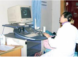
Wide-field ultrasound imaging technology, also known as ultra-wide field imaging, wide-field imaging or panoramic ultrasound imaging technology, is to obtain a series of two-dimensional slice images through the movement of the probe, and then use the computer reconstruction method to stitch this series of two-dimensional images into A cut-away image of a continuous super wide field of view. The main feature of wide-field ultrasound imaging technology is that it can provide better structural level and spatial relationship, clearly show the lesion location, size, range, internal echo and its adjacent, quantitatively and accurately measure the size and volume of the lesion or Tumors, which better display and extend the structure of the pipeline, have the main disadvantage of being disturbed by images of tissue or organ motion resulting in blurred images. Wide-field ultrasound imaging technology has been widely used in the diagnosis of muscle, blood vessel and peripheral nerve diseases of small organs such as chest and abdomen, obstetrics and gynecology, breast, thyroid, testis and limbs. A wide-field ultrasound image can completely display the entire breast, and the acquired image shape is the same as the natural shape of the breast. The breast anatomy is clear, the lesion features are clear, the contrast of different tissue structures is obvious, and the breast augmentation surgery filling material and pectoralis major muscle can be clearly displayed. The relationship between breast glands. A wide-field ultrasound image can also obtain the entire fetus, which is not available in conventional two-dimensional ultrasound, and even the complete structure including the placenta. For multiple pregnancy, fetal position judgment, evaluation of the amount and distribution of amniotic fluid, positioning of the placenta, measurement It has important value with grading and so on. In particular, the soft tissue of the limb trunk uses a high-frequency linear array probe for a wide range of rapid tomographic scans, and a wide and normal anatomical image of the skin, subcutaneous tissue, muscles, tendons, blood vessels, peripheral nerve trunks, and periosteum can be obtained. And the structural characteristics of each layer are clear at a glance. Wide-field ultrasound imaging technology has great development potential and good application prospects. It combines conventional real-time gray-scale and color Doppler ultrasound to make modern ultrasound diagnostic technology more perfect, and also lays the foundation for the research and application of ultrasound CT. The foundation.
3. Molecular imaging technology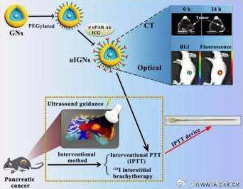
Molecular imaging is based on molecular imaging, based on molecular biology, to study and observe the occurrence of diseases at the molecular level, to develop pathophysiological changes and metabolic function changes, that is, to determine and describe living biological processes at the cellular and molecular levels. An imaging method. The terminology of molecular imaging first appeared in the mid to late 1990s and was officially proposed and applied by the National Cancer Institute in 1998. Molecular imaging differs from traditional imaging methods in that it reveals cellular and molecular abnormalities that cause disease in humans, rather than the final anatomical abnormalities caused by changes in cells or molecules. Clinically, because many diseases have obvious changes in function or cell molecules before pathological changes occur in organ tissues, cell molecular imaging technology can not only detect and determine diseases earlier, but also treat diseases. The effect directly evaluates the level of cells and molecules, thus establishing a new scientific understanding of the occurrence, development and healing of the disease. Ultrasound in molecular imaging through monoclonal antibodies, peptide molecules and other targeted microbubble contrast agents, can be used for cardiovascular, tumor and other targeted diagnosis, treatment of thrombosis, atherosclerotic plaque, and drug, gene delivery. Microbubbles and acoustically active substances can be used as targeting targets for ultrasound imaging, carrying targeting ligands, binding to living cells, for molecular imaging and therapy, targeting micro/nano bubbles to open a new frontier for molecular imaging . Molecular imaging is the result of multidisciplinary integration of molecular biology, biochemistry, nanotechnology, genetic engineering technology, data processing and image processing. It is also an inevitable trend of the revolutionary development and future development of modern medical imaging technology.
The company adheres to the management policy of "seeking truth and dedication" and a strict quality assurance system. In the new century, we will follow the tenet of "innovation, progress and rigor" and a forge ahead enterprise with "excellent quality and excellence". Spirit, the supreme product, add wings to your enterprise take off. Carefully do a good job of each product, the highest quality, and integrate a dedicated attitude, dedicated spirit, skilled practice, and good reputation into the details of the service.
High Voltage Connector,High Voltage Terminal Wire Connectors,High Voltage Terminal Connectors,High Voltage Terminal Connector Terminal
Sichuan Xinlian electronic science and technology Company , https://www.sztmlchs.com